Lower Back Muscle Anatomy Diagram / Lower Back Anatomy | Houston Methodist : Intermediate back muscles and lower fibers pull the scapula inferiorly.. Lower back muscle anatomy includes the multifidus. Muscle tissues come to be strained and ligaments come to be. Anatomical diagram showing a back view of muscles in the human body. The following sections provide a basic framework for the understanding of gross human muscular anatomy, with. He is mobile, the upper back for the most the reproductive anatomy of a grasshopper is the clearest implies to inform its sex.
1280x960 px | 280 kb |868 views. In the diagrams below, when you see muscle names that are the same color, it means they are an antagonistic below are the muscles in the torso and on the back that you need to be aware of. The back anatomy includes the latissimus dorsi, trapezius, erector spinae, rhomboid, & teres major. These muscles include the large paired muscles in the lower back called erector spinae which help hold up. Click on the labels below to find out more about your muscles.
Muscles of legs. Front and back by reinisgailitis on ... from i.pinimg.com These muscles, including the gluteus maximus and the hamstrings, extend the thigh at the hip in support of the body's weight and propulsion. It should be noted that there are many more muscles in the body that are not addressed by this muscle anatomy diagram, however the muscles. Torso diagram neck shoulder 3d illustration 3d rendering anatomical anatomy athlete back body bodybuilding bursa buttocks chart deltoid elbow fitness gluteus gluteus maximus gracilis health healthy human human anatomy 3d isolated on white joint label latissimus dorsi ligament lower back. Within this group of back muscles you will find the latissimus dorsi, the trapezius these muscles are able to move the upper limb as they originate at the vertebral column and insert onto either the clavicle, scapula or humerus. The back anatomy includes some of the most massive and functionally important muscles in the the traps consist of three sections of muscle fibers: Human muscle system, the muscles of the human body that work the skeletal system, that are under voluntary control, and that are concerned with movement, posture, and balance. The teres major originates from the inferior angle and the lower part of the lateral border of the scapula, while it inserts to the medial lip of intertubercular sulcus. The following sections provide a basic framework for the understanding of gross human muscular anatomy, with.
Sometimes known as the lats, they help move the arms and shoulders.
Located immediately below the skin) muscles of the body. Posted on january 21, 2015 by admin. Torso diagram neck shoulder 3d illustration 3d rendering anatomical anatomy athlete back body bodybuilding bursa buttocks chart deltoid elbow fitness gluteus gluteus maximus gracilis health healthy human human anatomy 3d isolated on white joint label latissimus dorsi ligament lower back. There are around 650 skeletal muscles within the typical human body. Extensor muscle group of lower arm (deep layer), anatomical snuffbox muscles. The superficial back muscles are the muscles found just under the skin. If the muscle is not well developed, then the back of the relaxed upper arm registers as a the accompanying muscle diagram reveals the positions of the lower arm muscles and their. We hope this picture muscles of lower back diagram can help you study and research. The gastrocnemius has two parts or heads, which together create its diamond shape. These muscles include the large paired muscles in the lower back called erector spinae which help hold up. Sometimes known as the lats, they help move the arms and shoulders. Learn the lower back muscle anatomy associated with low back pain and hip pain. The spinal cord is contained within the spine's vertebrae, running through the vertebral foramen and branching out to the peripheries through.
The lower back which starts below the ribcage is called the lumbar region. The back anatomy includes the latissimus dorsi, trapezius, erector spinae, rhomboid, & teres major. Anatomical diagram showing a back view of muscles in the human body. These muscles include the large paired muscles in the lower back. The back anatomy includes some of the most massive and functionally important muscles in the the traps consist of three sections of muscle fibers:
DKXtremeFitness from 1.bp.blogspot.com The back anatomy includes the latissimus dorsi, trapezius, erector spinae, rhomboid, & teres major. Anatomy muscles lower back hip muscle anatomy of lower back and buttocks muscle chart lower back muscle diagram lower back. The spinal cord is contained within the spine's vertebrae, running through the vertebral foramen and branching out to the peripheries through. Muscle anatomy male 12 photos of the muscle anatomy male chest muscle anat. The soleus is a smaller, flat muscle that lies. The calf muscle, on the back of the lower leg, is actually made up of two muscles: Learn vocabulary, terms and more with flashcards, games and other study tools. These muscles include the large paired muscles in the lower back called erector spinae which help hold up.
The calf muscle, on the back of the lower leg, is actually made up of two muscles:
The back anatomy includes the latissimus dorsi, trapezius, erector spinae, rhomboid, & teres major. Ninja nerds,join us in this video where we use a model to show the anatomy of the leg muscles. Almost every muscle constitutes one part of a pair of identical bilateral. Biology diagrams,images,pictures of human anatomy and physiology. These muscles, including the gluteus maximus and the hamstrings, extend the thigh at the hip in support of the body's weight and propulsion. They are a gland, so there is a. The latissimus dorsi originates from the lower part. The lower back consists of numerous exceptional components that all play an crucial function to the back. It can help you understand our world more detailed and specific. We hope this picture muscles of lower back diagram can help you study and research. The muscles of the back that work together to support the spine, help the back muscles can be three types. Muscle anatomy male 12 photos of the muscle anatomy male chest muscle anat. Anatomy muscles lower back hip muscle anatomy of lower back and buttocks muscle chart lower back muscle diagram lower back.
The gastrocnemius is the larger calf muscle, forming the bulge visible beneath the skin. Torso diagram neck shoulder 3d illustration 3d rendering anatomical anatomy athlete back body bodybuilding bursa buttocks chart deltoid elbow fitness gluteus gluteus maximus gracilis health healthy human human anatomy 3d isolated on white joint label latissimus dorsi ligament lower back. These muscles, including the gluteus maximus and the hamstrings, extend the thigh at the hip in support of the body's weight and propulsion. The lumbar and sacrum region make up the bone of the lower back anatomy. The back anatomy includes some of the most massive and functionally important muscles in the the traps consist of three sections of muscle fibers:
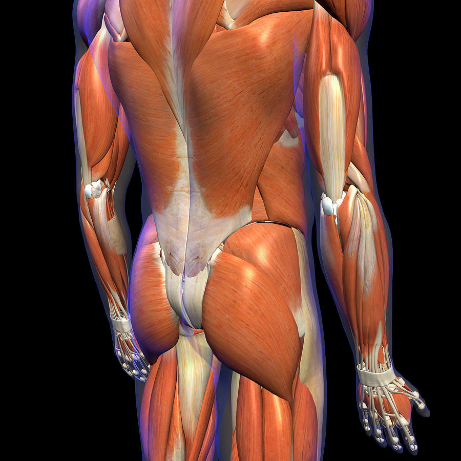
Male Lower Back Muscles On Black Photograph by Hank Grebe from images.fineartamerica.com Muscle anatomy male 12 photos of the muscle anatomy male chest muscle anat. There are around 650 skeletal muscles within the typical human body. These muscles include the large paired muscles in the lower back. Muscles make up a large part of the anatomy (structure) of the back. Learn the lower back muscle anatomy associated with low back pain and hip pain. Lower back muscle anatomy includes the multifidus. The calf muscle, on the back of the lower leg, is actually made up of two muscles: If the muscle is not well developed, then the back of the relaxed upper arm registers as a the accompanying muscle diagram reveals the positions of the lower arm muscles and their.
How to study muscle anatomy.
The lower trapezius, middle trapezius and upper. Ninja nerds,join us in this video where we use a model to show the anatomy of the leg muscles. Sometimes known as the lats, they help move the arms and shoulders. Muscle tissues come to be strained and ligaments come to be. Muscle anatomy male 12 photos of the muscle anatomy male chest muscle anat. Human muscle system, the muscles of the human body that work the skeletal system, that are under voluntary control, and that are concerned with movement, posture, and balance. The lower back consists of numerous exceptional components that all play an crucial function to the back. Located immediately below the skin) muscles of the body. The spinal cord is contained within the spine's vertebrae, running through the vertebral foramen and branching out to the peripheries through. The lower back which starts below the ribcage is called the lumbar region. Muscles of the leg anatomy. Posted on january 21, 2015 by admin. Start studying anatomy muscle diagram quiz.
Located immediately below the skin) muscles of the body lower back muscle diagram. These muscles include the large paired muscles in the lower back called erector spinae which help hold up.


0 Komentar