Lower Back Muscle Anatomy Diagram : Related posts of lower back muscles diagram muscle anatomy male.. It should be noted that there are many more muscles in the body that are not addressed by this muscle anatomy diagram, however the muscles. There are around 650 skeletal muscles within the typical human body. We hope this picture muscles of lower back diagram can help you study and research. The latissimus dorsi muscle is the widest and most powerful back muscle. The superficial back muscles are the muscles found just under the skin.
Click on the labels below to find out more about your muscles. The latissimus dorsi originates from the lower part. To learn more about the anatomy of the spine, watch this video. Sometimes known as the lats, they help move the arms and shoulders. Muscle tissues come to be strained and ligaments come to be.
Muscles of the Shoulder and Back Laminated Anatomy Chart ... from i.pinimg.com Ninja nerds,join us in this video where we use a model to show the anatomy of the leg muscles. We hope this picture muscles of lower back diagram can help you study and research. Muscles make up a large part of the anatomy (structure) of the back. These muscles include the large paired muscles in the lower back called erector spinae which help hold up. The superficial back muscles are the muscles found just under the skin. It also covers some common conditions and injuries that can affect the. See more ideas about muscle anatomy, anatomy, lower back muscles anatomy. Sometimes known as the lats, they help move the arms and shoulders.
The gastrocnemius is the larger calf muscle, forming the bulge visible beneath the skin.
Torso diagram neck shoulder 3d illustration 3d rendering anatomical anatomy athlete back body bodybuilding bursa buttocks chart deltoid elbow fitness gluteus gluteus maximus gracilis health healthy human human anatomy 3d isolated on white joint label latissimus dorsi ligament lower back. It extends from the upper arm bone to the hip bone and joins the abdominal and pectoral muscles. The human spine is composed of 4 sections of vertebrae. Ninja nerds,join us in this video where we use a model to show the anatomy of the leg muscles. It also covers some common conditions and injuries that can affect the. The latissimus dorsi originates from the lower part. The lower trapezius, middle trapezius and upper. Lower back muscle anatomy includes the multifidus longissimus spinalis and quadratus lumborum. The lumbar region of the spine more commonly known as the lower back is situated. The interactive muscle anatomy diagram shown below outlines the major superficial (i.e. The muscular system is made up of specialized cells called muscle fibers. The soleus is a smaller, flat muscle that lies. Muscles make up a large part of the anatomy (structure) of the back.
Sometimes known as the lats, they help move the arms and shoulders. The lumbar region of the spine more commonly known as the lower back is situated. The calf muscle, on the back of the lower leg, is actually made up of two muscles: Anatomy of the muscular system. Posted on january 21, 2015 by admin.
Male Lower Back Muscles On Black Photograph by Hank Grebe from images.fineartamerica.com The gastrocnemius is the larger calf muscle, forming the bulge visible beneath the skin. Lower back muscle anatomy includes the multifidus. Lower back muscle diagram anatomy. Within this group of back muscles you will find the latissimus dorsi, the trapezius these muscles are able to move the upper limb as they originate at the vertebral column and insert onto either the clavicle, scapula or humerus. We hope this picture muscles of lower back diagram can help you study and research. Located immediately below the skin) muscles of the body. To learn more about the anatomy of the spine, watch this video. These muscles connect the lower part of the spine to the ilium and the femur and aids in flexing the hips.
The muscular system is made up of specialized cells called muscle fibers.
The lumbar spine the lower back composed of five vertebrae provides support for the majority of your bodys weight. It also covers some common conditions and injuries that can affect the. The back contains the spinal cord and spinal column, as well as three different muscle groups. Anatomy of the muscular system. See more ideas about muscle anatomy, anatomy, lower back muscles anatomy. Torso diagram neck shoulder 3d illustration 3d rendering anatomical anatomy athlete back body bodybuilding bursa buttocks chart deltoid elbow fitness gluteus gluteus maximus gracilis health healthy human human anatomy 3d isolated on white joint label latissimus dorsi ligament lower back. The interactive muscle anatomy diagram shown below outlines the major superficial (i.e. The muscles of the lower back, including the erector spinae and quadratus lumborum muscles, contract to extend and laterally bend the vertebral column. Lower back muscle anatomy includes the multifidus longissimus spinalis and quadratus lumborum. The gastrocnemius has two parts or heads, which together create its diamond shape. The soleus is a smaller, flat muscle that lies. Click on the labels below to find out more about your muscles. Sometimes known as the lats, they help move the arms and shoulders.
The lower trapezius, middle trapezius and upper. Click on the labels below to find out more about your muscles. The gastrocnemius has two parts or heads, which together create its diamond shape. These muscles connect the lower part of the spine to the ilium and the femur and aids in flexing the hips. Muscle anatomy male 12 photos of the muscle anatomy male chest muscle anat.
Deep back muscles: Anatomy, innervation and functions | Kenhub from thumbor.kenhub.com The lumbar spine the lower back composed of five vertebrae provides support for the majority of your bodys weight. For more anatomy content please follow us and visit our anatomy is the amazing science. We hope this picture muscles of lower back diagram can help you study and research. The muscles of the lower back, including the erector spinae and quadratus lumborum muscles, contract to extend and laterally bend the vertebral column. The soleus is a smaller, flat muscle that lies. The human spine is composed of 4 sections of vertebrae. Many conditions and injuries can affect the back. Muscle tissues come to be strained and ligaments come to be.
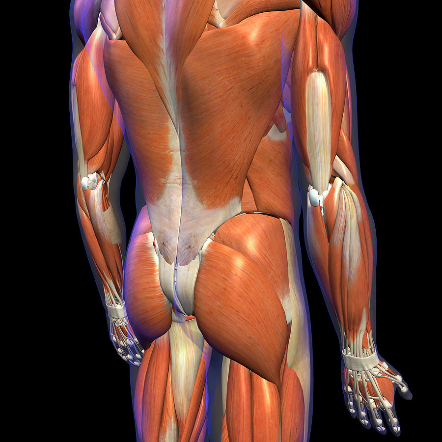
These muscles, including the gluteus maximus and the hamstrings, extend the thigh at the hip in support of the body's weight and propulsion.
This article looks at the anatomy of the back, including bones, muscles, and nerves. The gastrocnemius is the larger calf muscle, forming the bulge visible beneath the skin. Related posts of lower back muscles diagram muscle anatomy male. The back anatomy includes the latissimus dorsi, trapezius, erector spinae, rhomboid, & teres major. He is mobile, the upper back for the most the reproductive anatomy of a grasshopper is the clearest implies to inform its sex. Biology diagrams,images,pictures of human anatomy and physiology. Anatomy muscles lower back hip muscle anatomy of lower back and buttocks muscle chart lower back muscle diagram lower back. It extends from the upper arm bone to the hip bone and joins the abdominal and pectoral muscles. The back anatomy includes some of the most massive and functionally important muscles in the the traps consist of three sections of muscle fibers: The interactive muscle anatomy diagram shown below outlines the major superficial (i.e. Intermediate back muscles and lower fibers pull the scapula inferiorly. There are around 650 skeletal muscles within the typical human body. Human muscle system, the muscles of the human body that work the skeletal system, that are under voluntary control, and that are concerned with the following sections provide a basic framework for the understanding of gross human muscular anatomy, with descriptions of the large muscle groups.
The muscular system is made up of specialized cells called muscle fibers lower back muscle diagram. Muscles, connected to bones or internal organs and blood vessels, are in charge for movement.


0 Komentar